 English
  Nederlands
  French
  Deutsch
 Perfusion can be visualized during surgery with help of the Quest Spectrum®. During an open or laparoscopic procedure, patients are injected with Indocyanine Green (ICG) which binds to plasma proteins within the vascular system. This tracer flows throughout the cardiovascular system including to the anatomy of interest.
Breast Reconstruction
Skin flap necrosis up to 14% in flap reconstruction
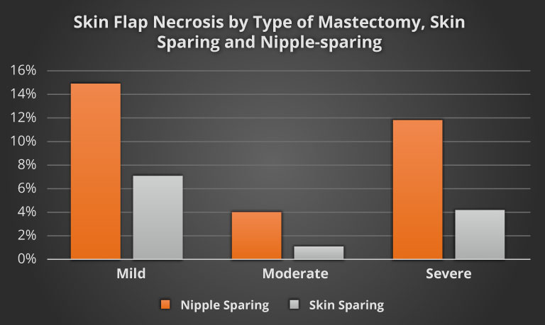
Matsen C, et al. Skin Flap Necrosis After Mastectomy With Reconstruction: A Prospective Study. Ann Surg Oncol. 2016 Jan: 23(1): 257-264.
FGS helps lower complication rates by up to 26%
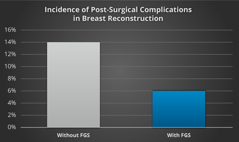
Duggal CS. An Outcome Analysis of Intraoperative Angiography for Postmastectomy Breast Reconstruction. Aesthetic Surgery Journal. 2014; 34(1):61-5.
Colorectal Surgeries
Anastomotic leaks up to 19% in GI tract
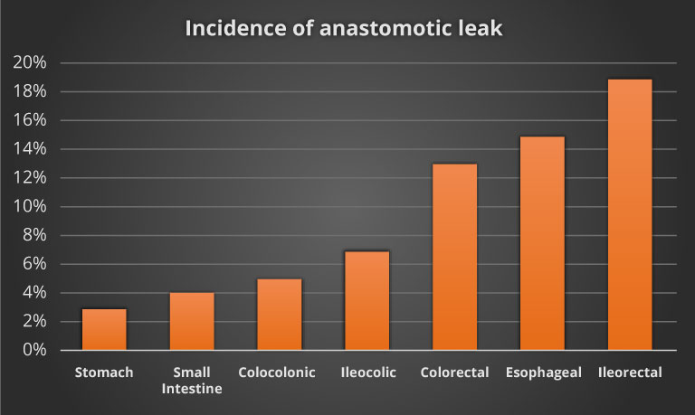
Phillips B. Reducing gastrointestinal anastomotic leak rates: review of challenges and solutions. Open Access Surgery. 2016; 9:5-14
Anastomotic leaks rates reduced by up to 55%
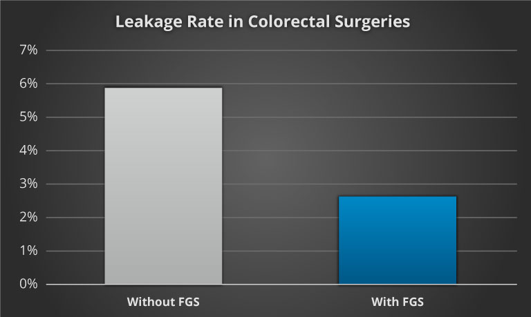
Ris F, Liot E, Buchs NC, et al. Multicenter phase II trial of near-infrared imaging in elective colorectal surgery. Br J Surg. 2018;105(10):1359-1367. doi:10.1002/bjs.10844
Combination of images
When the Quest Spectrum is used to simultaneously record the visible and near-infrared images, the near-infrared images reveal the location of the fluorescent tracer, while the visible images show the anatomy of interest. When these images are combined one can visualize what anatomy is perfused.
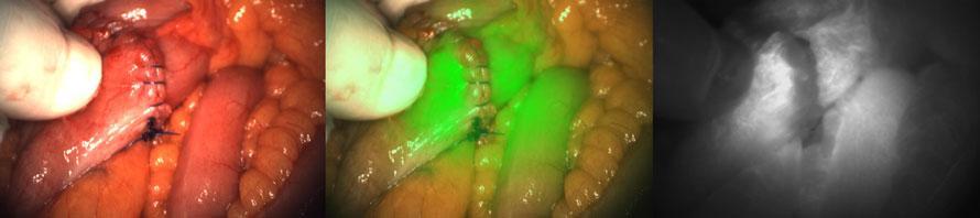
The perfusion of the colon during a colorectal anastomosis procedure is shown above using the Quest Spectrum.
- The left image shows an image similar to what the surgeon sees with the naked eye. This image shows a closed suture without any leakages. One cannot estimate the perfusion of the suture based on this image.
- The right image shows the response of the fluorescent tracer that is used during this procedure. This image shows that the intensity of the fluorescent response is lower at the center of the image compared to the surrounding tissue, indicating less perfusion.
- The image in the middle shows the overlay between the visible image and the fluorescent image. This image allows the surgeon to assess perfusion in the anatomic structures of concern.
Estimating perfusion in these procedures can enable the surgeon to reduce complications, late effects, morbidity and mortality.
Links to supporting literature:
Clinical role of fluorescence imaging in colorectal surgery – a review
Mazrahi & Wexner
Expert Rev Med Devices. 2017 Jan; 14(1):75-82.
